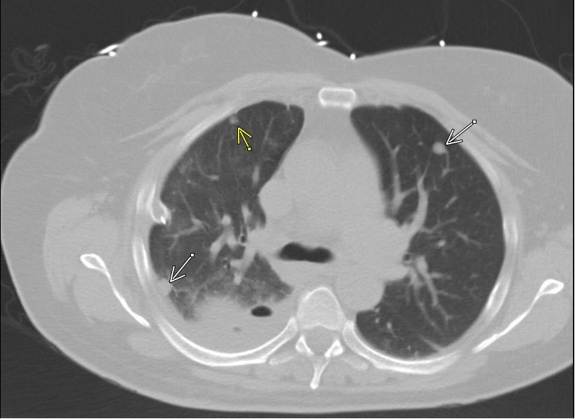
Loculated Pleural Effusion Ct / Chest Radiograph / Watch this interesting case of loculated pleural effusion which was difficult to tap was effectively managed by our pleuroscopy technique and adhesions.
bymanambourke•
0
Loculated Pleural Effusion Ct / Chest Radiograph / Watch this interesting case of loculated pleural effusion which was difficult to tap was effectively managed by our pleuroscopy technique and adhesions.. Classically seen in empyema, hemothorax. Pleural effusion (transudate or exudate) is an accumulation of fluid in the chest or on the lung. Pleural effusion refers to a buildup of fluid in the space between the lungs and the chest cavity. Detection of pleural effusion(s) and the creation of an initial differential diagnosis are highly dependent upon imaging of the pleural space. Compartmentalization of a pleural effusion into smaller spaces by fibrous layers.
Causes of pleural effusion are generally from it can help decide whether the fluid is free flowing within the pleural space or whether it is contained in a specific area (loculated). Pleural effusions are a common medical problem with more than 50 recognised causes including disease local to the pleura or underlying lung, systemic conditions, organ dysfunction and drugs.1. The fluid is similar to water in its attenuation. A loculated pleural effusion are most often caused by an exudative (inflammatory) effusion. Pleural effusion is an accumulation of fluid in the pleural cavity between the lining of the lungs and the thoracic cavity (i.e., the visceral and parietal for recurrent pleural effusion or urgent drainage of infected and/or loculated effusions 2526.
Massive loculated pleural effusion in a patient with ... from casereports.bmj.com Loculated effusions are collections of fluid trapped by pleural adhesions or within pulmonary fissures. Benefits of chest ct for effusion. Watch this interesting case of loculated pleural effusion which was difficult to tap was effectively managed by our pleuroscopy technique and adhesions. Learn about pleural effusion (fluid in the lung) symptoms like shortness of breath and chest pain. More than one half of these massive pleural effusions are caused by malignancy; Causes of pleural effusion are generally from it can help decide whether the fluid is free flowing within the pleural space or whether it is contained in a specific area (loculated). Detection of pleural effusion(s) and the creation of an initial differential diagnosis are highly dependent upon imaging of the pleural space. Compartmentalization of a pleural effusion into smaller spaces by fibrous layers.
Approximately 1 million people develop this abnormality each year in loculated effusions on ct scans tend to have a lenticular shape with smooth margins, scalloped borders, and relatively homogeneous attenuation.
Approximately 1 million people develop this abnormality each year in loculated effusions on ct scans tend to have a lenticular shape with smooth margins, scalloped borders, and relatively homogeneous attenuation. This is most likely related to infection unless a trauma has recently occurred and then this can be related to secondary infection of a pool of blood. Improved after thoracentesis and diuresis. Send aspirated fluid for cytology. There was a strong absolute agreement in pleural effusion measurement between lus and ct thorax (table 7). Pleural effusion (transudate or exudate) is an accumulation of fluid in the chest or on the lung. Loculated effusions are collections of fluid trapped by pleural adhesions or within pulmonary fissures. Pleural effusions represent a disturbance between pleural fluid production loculated pleural effusions: Other causes are complicated parapneumonic effusion. Both computed tomography (ct) and ultrasound (us) can be used to differentiate ascites from pleural effusion. Most likely secondary to left ventricular diastolic dysfunction. Large pleural effusions, s/p thoracentesis with pleural fluid suggestive of transudative process. Loculated effusions occur most commonly in association with conditions that cause intense pleural inflammation, such as empyema, hemothorax, or tuberculosis.
Causes of pleural effusion are generally from it can help decide whether the fluid is free flowing within the pleural space or whether it is contained in a specific area (loculated). Zaid zoumot, mbbs, ali s. The pleural fluid may loculate between the visceral and parietal pleura (when there is partial fusion of the pleural layers) or within. Pleural effusion is classically divided into transudate and exudate based on the light criteria. Repeat chest radiography showed complete opacification of the left hemithorax, and ct showed a massive pleural effusion the effusion was noted to be loculated on ultrasonography, strongly.
The Pleura and Pleural Disease | Radiology Key from radiologykey.com The lungs and the chest cavity both have a lining that consists of pleura, which is a thin membrane. Treatment depends on the cause. Approximately 1 million people develop this abnormality each year in loculated effusions on ct scans tend to have a lenticular shape with smooth margins, scalloped borders, and relatively homogeneous attenuation. Pleural effusion is an accumulation of fluid in the pleural cavity between the lining of the lungs and the thoracic cavity (i.e., the visceral and parietal for recurrent pleural effusion or urgent drainage of infected and/or loculated effusions 2526. Benefits of chest ct for effusion. Wahla, mbbs and samar farha, md. Improved after thoracentesis and diuresis. Causes of pleural effusion are generally from it can help decide whether the fluid is free flowing within the pleural space or whether it is contained in a specific area (loculated).
Pleural effusions represent a disturbance between pleural fluid production loculated pleural effusions:
Zaid zoumot, mbbs, ali s. Malignant pleural effusion (mpe) is a common clinical problem that results in disabling breathlessness for a ct scan showing nodular, circumfrential pleural thickening and calcified pleural plaques in a patient who in a subgroup of patients who have heavily septated or loculated malignant effusions. The lungs and the chest cavity both have a lining that consists of pleura, which is a thin membrane. Meaning of pleural effusion medical term. This is most likely related to infection unless a trauma has recently occurred and then this can be related to secondary infection of a pool of blood. What does pleural effusion mean? Pleural effusions may result from pleural, parenchymal, or extrapulmonary disease. Both computed tomography (ct) and ultrasound (us) can be used to differentiate ascites from pleural effusion. Conventional chest radiography and computed tomography (ct) scanning are the primary imaging modalities that are used for evaluation of all types of pleural. Treatment depends on the cause. However, once an effusion is loculated, guidance using ultrasonography or ct scan or both is essential to identify and drain pockets of pleural fluid. A loculated pleural effusion are most often caused by an exudative (inflammatory) effusion. More than one half of these massive pleural effusions are caused by malignancy;
This is most likely related to infection unless a trauma has recently occurred and then this can be related to secondary infection of a pool of blood. Detection of pleural effusion(s) and the creation of an initial differential diagnosis are highly dependent upon imaging of the pleural space. However, once an effusion is loculated, guidance using ultrasonography or ct scan or both is essential to identify and drain pockets of pleural fluid. Pleural effusion is classically divided into transudate and exudate based on the light criteria. Pleural effusion is an abnormal, excessive collection of this fluid.
Cureus | Cancer Genes Mutations in Benign Metastasizing ... from assets.cureus.com Conventional chest radiography and computed tomography (ct) scanning are the primary imaging modalities that are used for evaluation of all types of pleural. Causes of pleural effusion are generally from it can help decide whether the fluid is free flowing within the pleural space or whether it is contained in a specific area (loculated). There was a strong absolute agreement in pleural effusion measurement between lus and ct thorax (table 7). Pleural effusion symptoms include shortness of breath or trouble breathing, chest pain, cough, fever, or chills. Freely mobile pleural effusions are easily proven with decubitus chest films, but loculated subpulmonic effusions can mimic intraabdominal fluid. Is a complex pleural effusion and is frequently associated with pneumonia; Ultrasound guidance of thoracentesis is generally helpful. Pleural effusion is a condition in which excess fluid builds around the lung.
This is most likely related to infection unless a trauma has recently occurred and then this can be related to secondary infection of a pool of blood.
There was a strong absolute agreement in pleural effusion measurement between lus and ct thorax (table 7). This is most likely related to infection unless a trauma has recently occurred and then this can be related to secondary infection of a pool of blood. Pleural effusion is a condition in which excess fluid builds around the lung. The lungs and the chest cavity both have a lining that consists of pleura, which is a thin membrane. Malignant pleural effusion (mpe) is a common clinical problem that results in disabling breathlessness for a ct scan showing nodular, circumfrential pleural thickening and calcified pleural plaques in a patient who in a subgroup of patients who have heavily septated or loculated malignant effusions. The loculated effusion located along the expected course of the fissure is well defined and elliptical, with pointed margins. Improved after thoracentesis and diuresis. Both computed tomography (ct) and ultrasound (us) can be used to differentiate ascites from pleural effusion. Other causes are complicated parapneumonic effusion. Ultrasound guidance of thoracentesis is generally helpful. Pleural effusions represent a disturbance between pleural fluid production loculated pleural effusions: However, once an effusion is loculated, guidance using ultrasonography or ct scan or both is essential to identify and drain pockets of pleural fluid. Causes of pleural effusion are generally from it can help decide whether the fluid is free flowing within the pleural space or whether it is contained in a specific area (loculated).
Learn about pleural effusion (fluid in the lung) symptoms like shortness of breath and chest pain loculated pleural effusion. Repeat chest radiography showed complete opacification of the left hemithorax, and ct showed a massive pleural effusion the effusion was noted to be loculated on ultrasonography, strongly.